Find hidden anatomy
Focused light and magnification help identify small, calcified or additional canal openings that can be difficult to see unaided.
 Book a consultation
Microscopic endodontics
A root canal is measured in millimetres and carried out inside a space that is narrow, dark and often complex. A dental operating microscope brings focused illumination and magnification directly into that space, giving the clinician greater visual control during treatment.
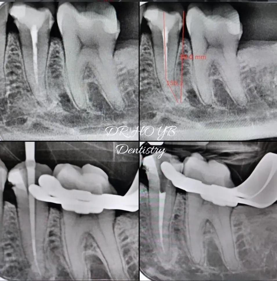
What is microscopic endodontics?
Root canal treatment removes inflamed or infected tissue from inside a tooth. The canal system is then cleaned, disinfected, filled and sealed so the natural tooth can remain in function.
The operating microscope does not change the biological principles of treatment. It improves how clearly the clinician can see the access cavity, canal openings and fine internal details while carrying out those principles.
This is especially valuable when canals are calcified, unusually curved, previously treated or hidden beneath old restorative material.
Benefits of magnification
Focused light and magnification help identify small, calcified or additional canal openings that can be difficult to see unaided.
A clearer view gives the clinician finer visual control when opening the tooth, negotiating canals and working around delicate structures.
Seeing the working field clearly can support a more conservative access—removing what is necessary while protecting sound tooth tissue where possible.
The microscope is particularly useful for retreatment, obstructions, ledges, perforations, separated instruments and complex root anatomy.
When does it help?
A careful sequence
The microscope is one part of a complete endodontic workflow. Each stage contributes to the result and the long-term outlook of the tooth.
We assess your symptoms, tooth, existing restoration and radiographs. A 3D CBCT scan may be recommended when the anatomy or source of infection needs further investigation.
Local anaesthetic is given and a rubber dam isolates the tooth, helping keep the treatment field clean and protecting the airway.
Magnification and focused light help the clinician work through a controlled opening and search systematically for the canal entrances.
The canals are carefully negotiated, measured, shaped and disinfected. Complex anatomy may require additional time or visits.
Once the canal system is ready, it is filled and sealed to reduce the space in which bacteria can persist or re-enter.
A strong final restoration—often an onlay or crown for a heavily weakened back tooth—helps protect the tooth from leakage and fracture.
Long-term tooth retention
Greater visibility can support more complete treatment, precise problem-solving and preservation of tooth structure. These advantages may improve the conditions for healing and help a root-canal-treated tooth remain useful for longer.
However, magnification alone cannot guarantee success. The prognosis also depends on the original infection, root anatomy, cracks, the amount of healthy tooth remaining, the quality and timing of the final restoration, biting forces, oral hygiene and follow-up care.
Clinical work
Clinical images and videos are shared for education. Every tooth has different anatomy, challenges and healing potential; individual results vary.
Microscope-assisted visualisation supported the search for and treatment of a complex four-canal system.
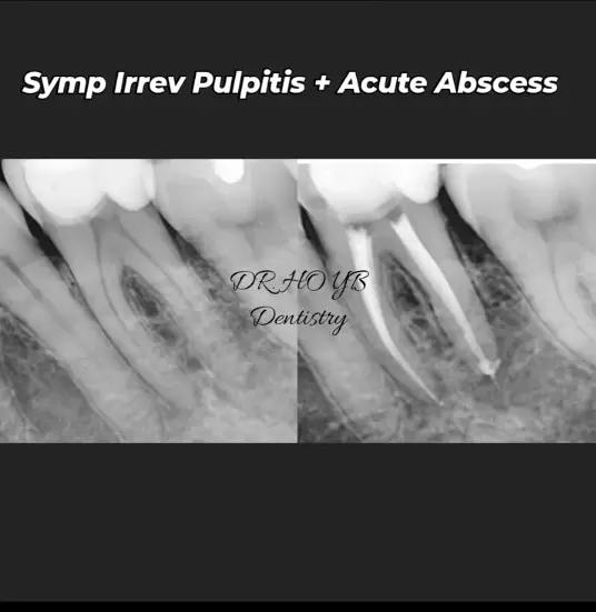
Diagnostic and post-treatment radiographs documenting endodontic management of an acutely infected tooth.
Inside the treatment
Short clinical videos demonstrating magnification-assisted treatment and retreatment.

Your endodontic clinician
Dr Ho is an honours graduate of Trinity College Dublin, a Member of the Faculty of Dentistry of the Royal College of Surgeons in Ireland and a recipient of the John Smith Medal from the Royal College of Surgeons of Edinburgh.
Her clinical focus is endodontics, with an emphasis on careful diagnosis, microscope-assisted precision and helping patients preserve their natural teeth whenever appropriate.
Consult Dr Ho →Toothache, swelling or a previously treated tooth?
A consultation and appropriate imaging are needed before we can advise whether root canal treatment, retreatment or another option is suitable.
WhatsApp our team →